Skip to product information
1 of 6

PayPal, credit cards. Download editable-PDF and invoice in 1 second!

YY/T 1465.6-2019 English PDF (YYT1465.6-2019)

YY/T 1465.6-2019 English PDF (YYT1465.6-2019)

Regular price $185.00 USD
Regular price Sale price $185.00 USD
Sale Sold out
Shipping calculated at checkout.
Delivery: 3 seconds (Download full-editable-PDF + Invoice).
Quotation: Click YY/T 1465.6-2019>>Add to cart>>Quote
Editable-PDF Preview (Reload if blank, scroll for next page)

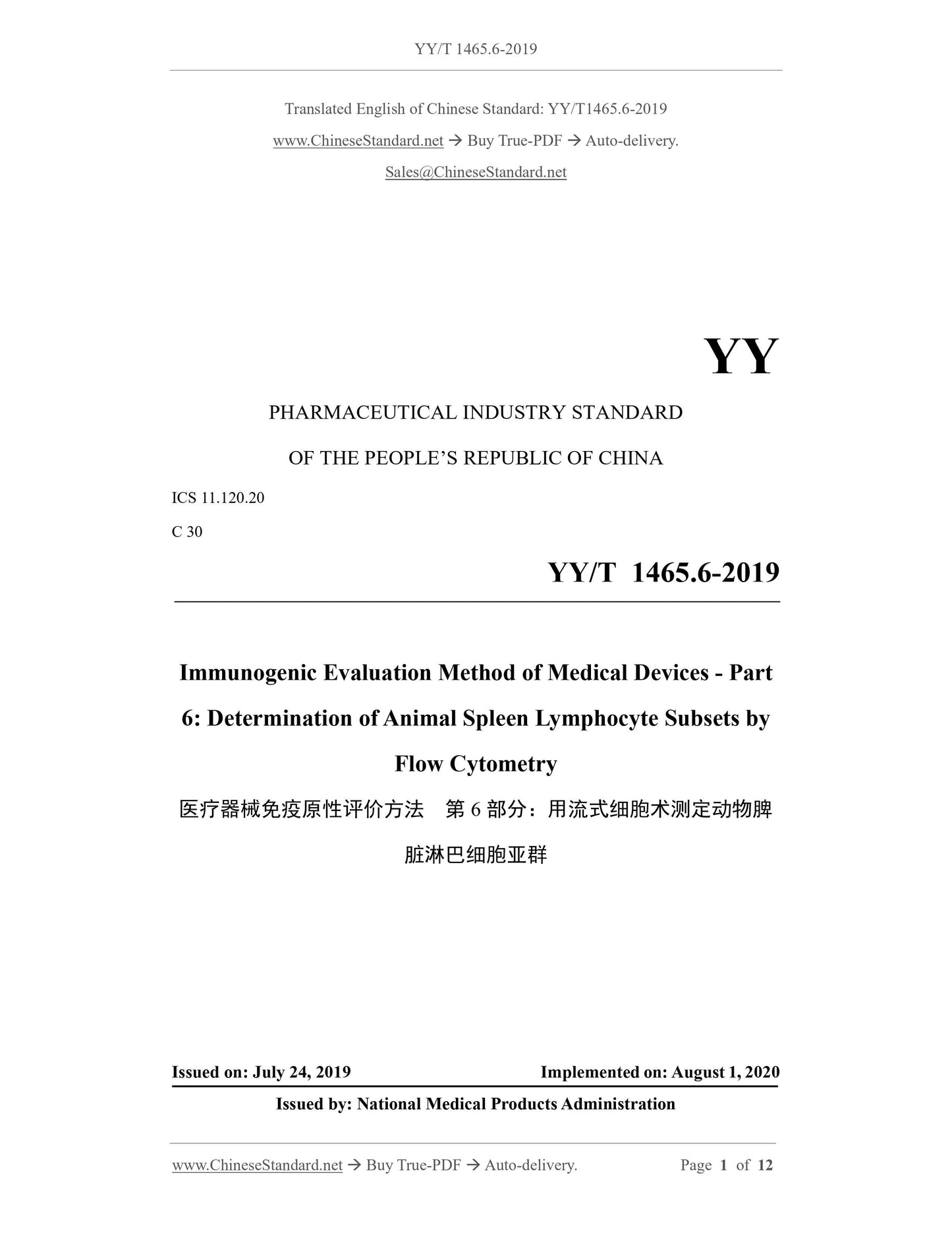
YY/T 1465.6-2019: Immunogenic evaluation method of medical devices - Part 6: Determination of animal spleen lymphocyte subsets by flow cytometry
This Part of YY/T 1465 specifies the method for the determination of animal spleen lymphocyte subsets by flow cytometry. This Part is applicable to the evaluation of immune responses induced by medical devices / materials.
YY/T 1465.6-2019
Immunogenic evaluation method of medical devices - Part 6. Determination of animal spleen lymphocyte subsets by flow cytometry ICS 11.120.20
C30
People's Republic of China Pharmaceutical Industry Standard
Medical device immunogenicity evaluation method
Part 6. Determination by flow cytometry
Animal spleen lymphocyte subset
Immunogenicevaluationmethodofmedicaldevices-
Part 6.Determinationofanimalspleenlymphocytesubsetsbyflowcytometry
Published on.2019-07-24
2020-08-01 implementation
State Drug Administration issued
Foreword
YY/T 1465 "Method for Evaluating Immunogenicity of Medical Devices" is divided into the following sections. --- Part 1. In vitro T lymphocyte transformation test;
--- Part 2. Determination of serum immunoglobulin and complement components (ELISA method); --- Part 3. Determination of plaque forming cells by agar solid phase method; --- Part 4. Half-in vivo method for phagocytosis of chicken red blood cells by mouse peritoneal macrophages; --- Part 5. Determination of α-Gal antigen clearance in animal-derived medical devices using the M86 antibody; --- Part 6. Determination of spleen lymphocyte subsets in animals by flow cytometry. This part is part 6 of YY/T 1465.
This part is drafted in accordance with the rules given in GB/T 1.1-2009. Please note that some of the contents of this document may involve patents. The issuing organization of this document is not responsible for identifying these patents. This part is proposed by the State Drug Administration.
This part is under the jurisdiction of the National Technical Committee for Standardization of Medical Device Biology Evaluation (SAC/TC248). This section drafted by. Shandong Province Medical Device Product Quality Inspection Center, Beijing Medical Device Inspection Institute. Drafters of this section. Hou Li, Sun Xiaoxia, Dai Zhengning, He Xueying, Wang Rui. introduction
The immune response is an important defense mechanism of the body. As an exogenous substance, medical devices pass through various ways after contact with the human body. The path affects the immune response of the body's immune system. Especially for animal-derived medical products, allogeneic products and tissue engineering medical products Wait. Although the interaction of medical devices/materials with the immune system may produce different immune responses, they can be broadly classified into two types. That is, humoral immune response and cell-mediated immune response. At present, it is not possible to determine the immune response to the host caused by medical devices or materials. It is beneficial or harmful, so it is very important to apply medical devices/materials for immune response research to obtain relevant information. Guidance on the possible immune response and potential immunotoxicity of medical devices in contact with humans is given in GB/T 16886.20. However, there is a lack of specific test methods. The YY/T 1465 series of standards is expected to provide specific test methods for the implementation of GB/T 16886.20. This part of YY/T 1465 provides a method for the determination of spleen lymphocyte subsets in experimental animals by flow cytometry for medical devices/materials. Provide specific test methods for the body's immune response potential, which can be used as an option in the immunotoxicology test in GB/T 16886.20. The method standard chosen. Other methods approved for use may also be employed. Medical device immunogenicity evaluation method
Part 6. Determination by flow cytometry
Animal spleen lymphocyte subset
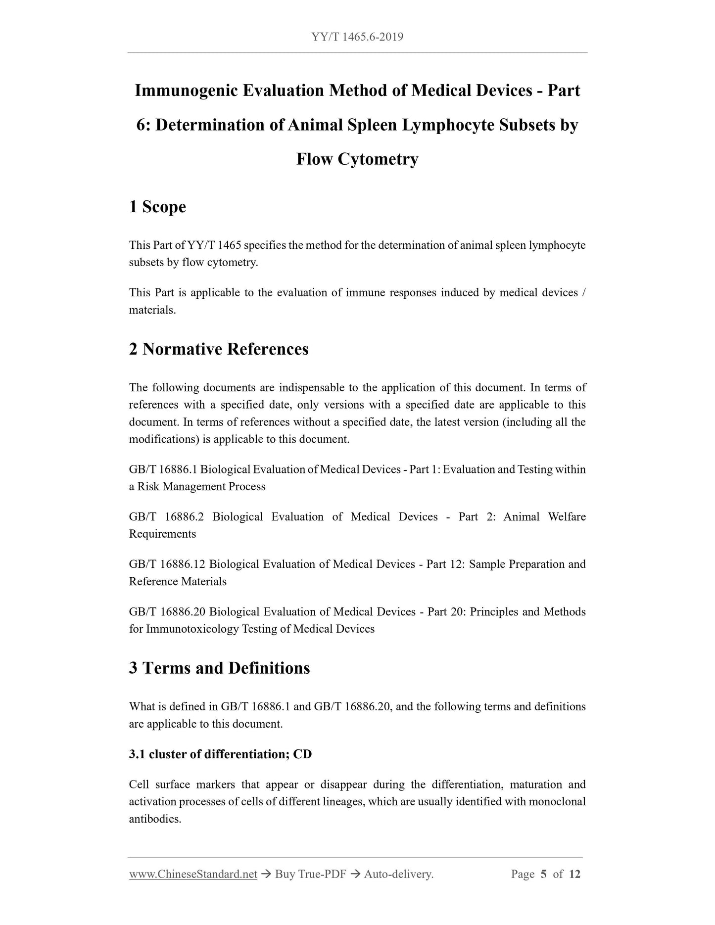
1 Scope
This section of YY/T 1465 specifies methods for determining lymphocyte subsets in spleen of animals by flow cytometry. This section applies to the evaluation of immune responses induced by medical devices/materials. 2 Normative references
The following documents are indispensable for the application of this document. For dated references, only dated versions apply to this article. Pieces. For undated references, the latest edition (including all amendments) applies to this document. GB/T 16886.1 Biological evaluation of medical devices - Part 1. Evaluation and testing in the process of risk management GB/T 16886.2 Biological evaluation of medical devices - Part 2. Animal welfare requirements GB/T 16886.12 Biological evaluation of medical devices - Part 12. Sample preparation and reference materials GB/T 16886.20 Biological evaluation of medical devices - Part 20. Principles and methods for immunological toxicology of medical devices 3 Terms and definitions
GB/T 16886.1, GB/T 16886.20 and the following terms and definitions apply to this document. 3.1
Differentiation antigen clusterofdifferentiation; CD
Cell surface markers that appear or disappear during differentiation, maturation, and activation of different lineage cells, usually identified by monoclonal antibodies. 3.2
Forward scattered light forwardanglelightscatter; FALs/FSC/Fs
The low angle optical signal collected by the optical detector directly in front of the incident light is related to the size and surface area of the cells or particles. 3.3
Side scattered light sidescatter; SSC
The optical signal scattered by the optical detector at the right angle of the incident light, complex with the internal and surface structure of the cell or particle The degree is related, such as cytoplasmic granularity, membrane irregularity and nuclear shape. 3.4
Fluorescence intensity
The amount of fluorescein bound to the cells or particles. The larger the number of channels of the fluorescent signal, the stronger the fluorescence intensity. In the right conditions The fluorescence intensity is related to the number of sites in which a single cell and a particular fluorescein bind. 4 Abbreviations
The following abbreviations apply to this document.
View full details