1
/
of
10
www.ChineseStandard.us -- Field Test Asia Pte. Ltd.
GB/T 39104.1-2020 English PDF (GB/T39104.1-2020)
Regular price
$320.00
Regular price
Sale price
$320.00
Unit price
/
per
Shipping calculated at checkout.
Couldn't load pickup availability
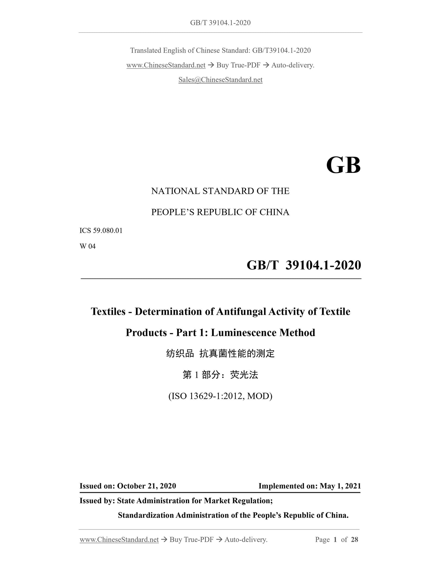
GB/T 39104.1-2020: Textiles - Determination of antifungal activity of textile products - Part 1: Luminescence method
Delivery: 9 seconds. Download (& Email) true-PDF + Invoice.
Get Quotation: Click GB/T 39104.1-2020 (Self-service in 1-minute)
Historical versions (Master-website): GB/T 39104.1-2020
Preview True-PDF (Reload/Scroll-down if blank)
GB/T 39104.1-2020
GB
NATIONAL STANDARD OF THE
PEOPLE’S REPUBLIC OF CHINA
ICS 59.080.01
W 04
Textiles - Determination of Antifungal Activity of Textile
Products - Part 1: Luminescence Method
(ISO 13629-1:2012, MOD)
ISSUED ON: OCTOBER 21, 2020
IMPLEMENTED ON: MAY 1, 2021
Issued by: State Administration for Market Regulation;
Standardization Administration of the People’s Republic of China.
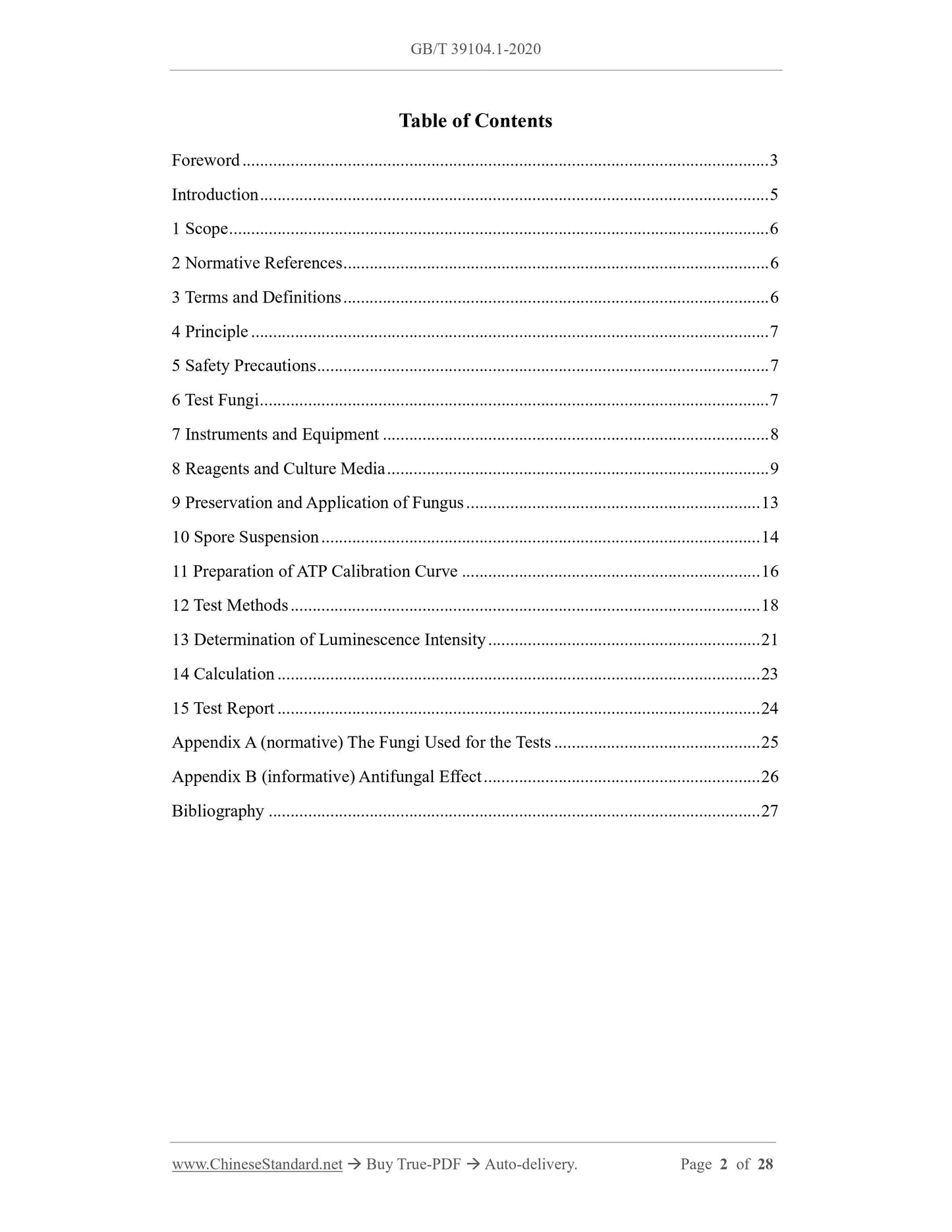
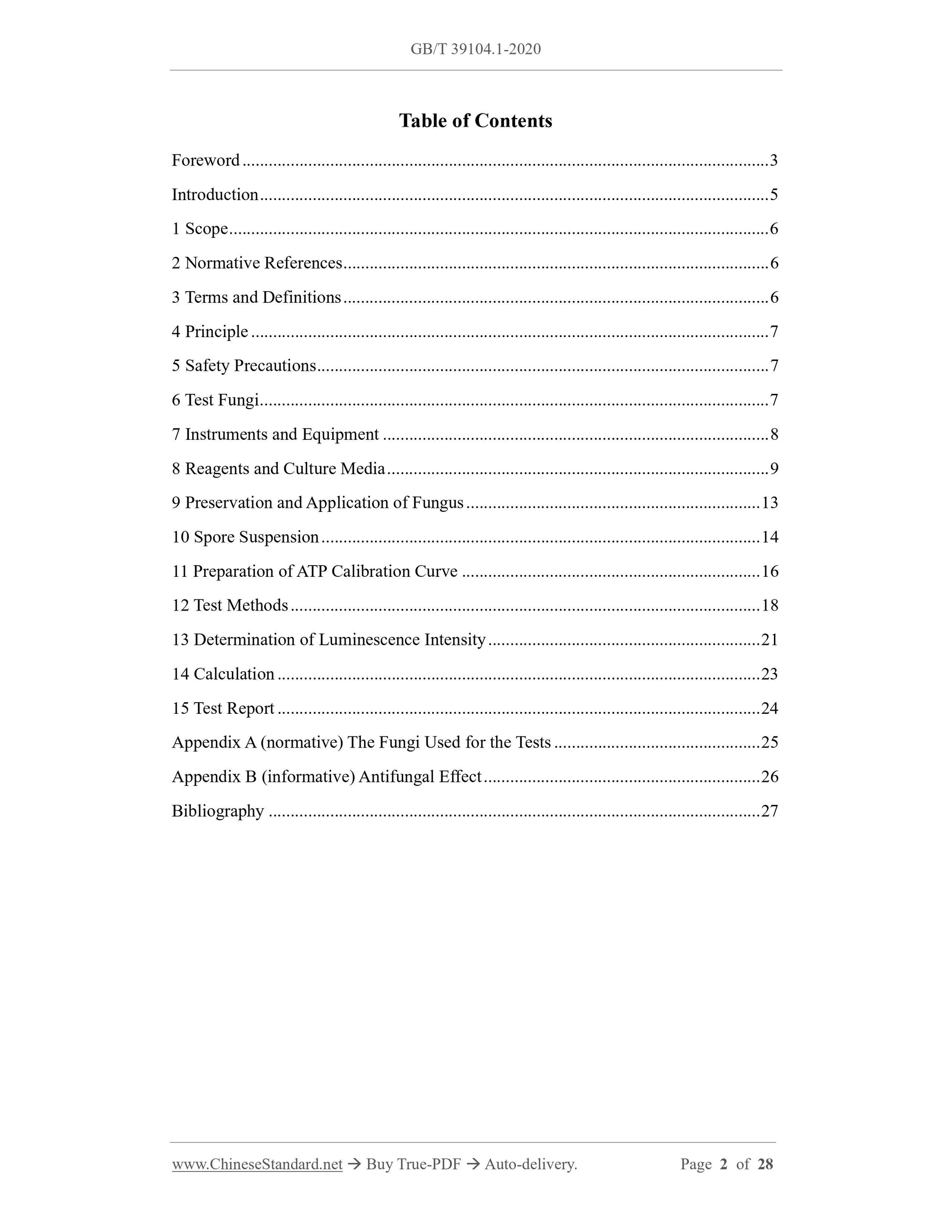
Table of Contents
Foreword ... 3
Introduction ... 5
1 Scope ... 6
2 Normative References ... 6
3 Terms and Definitions ... 6
4 Principle ... 7
5 Safety Precautions ... 7
6 Test Fungi ... 7
7 Instruments and Equipment ... 8
8 Reagents and Culture Media ... 9
9 Preservation and Application of Fungus ... 13
10 Spore Suspension ... 14
11 Preparation of ATP Calibration Curve ... 16
12 Test Methods ... 18
13 Determination of Luminescence Intensity ... 21
14 Calculation ... 23
15 Test Report ... 24
Appendix A (normative) The Fungi Used for the Tests ... 25
Appendix B (informative) Antifungal Effect ... 26
Bibliography ... 27
Textiles - Determination of Antifungal Activity of Textile
Products - Part 1: Luminescence Method
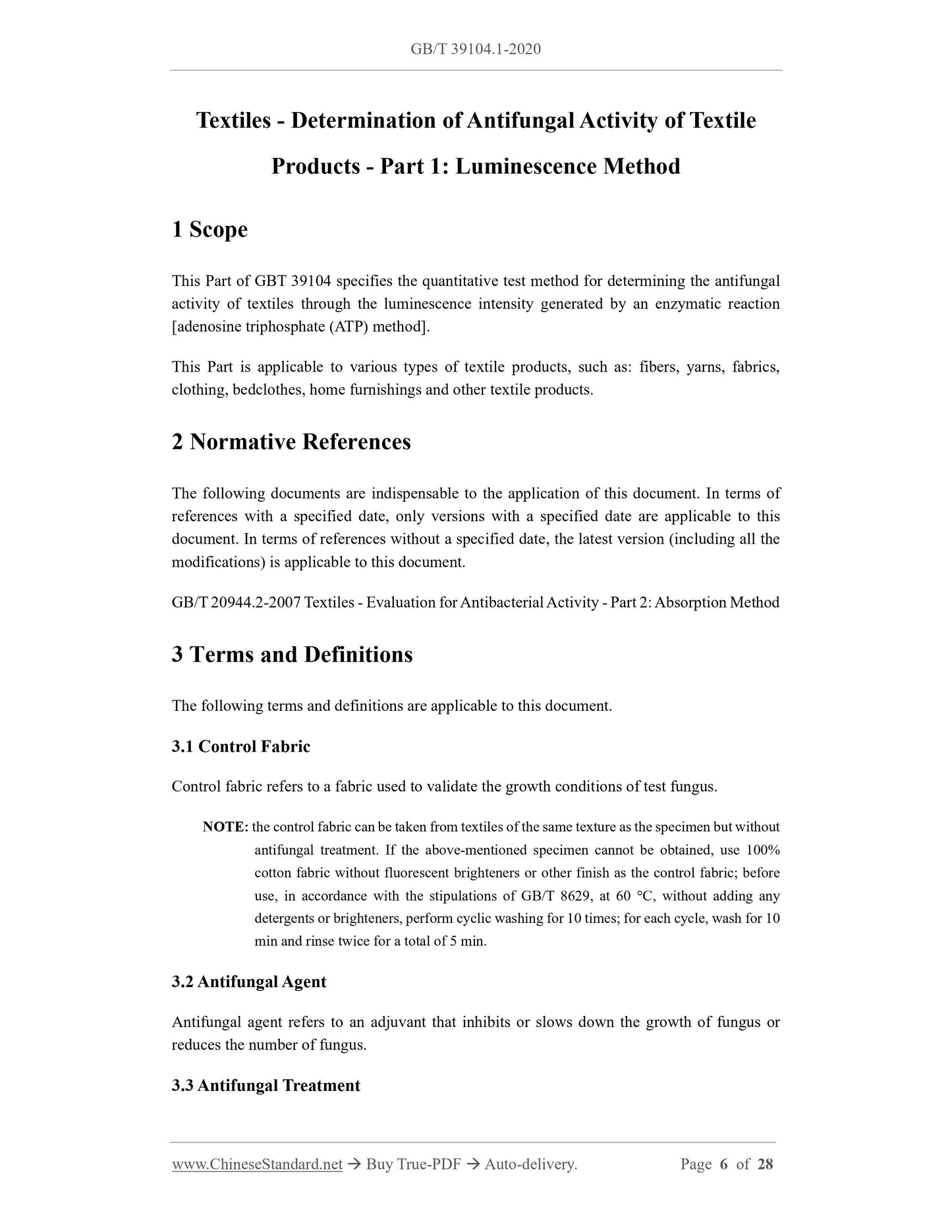
1 Scope
This Part of GBT 39104 specifies the quantitative test method for determining the antifungal
activity of textiles through the luminescence intensity generated by an enzymatic reaction
[adenosine triphosphate (ATP) method].
This Part is applicable to various types of textile products, such as: fibers, yarns, fabrics,
clothing, bedclothes, home furnishings and other textile products.
2 Normative References
The following documents are indispensable to the application of this document. In terms of
references with a specified date, only versions with a specified date are applicable to this
document. In terms of references without a specified date, the latest version (including all the
modifications) is applicable to this document.
GB/T 20944.2-2007 Textiles - Evaluation for Antibacterial Activity - Part 2: Absorption Method
3 Terms and Definitions
The following terms and definitions are applicable to this document.
3.1 Control Fabric
Control fabric refers to a fabric used to validate the growth conditions of test fungus.
NOTE: the control fabric can be taken from textiles of the same texture as the specimen but without
antifungal treatment. If the above-mentioned specimen cannot be obtained, use 100%
cotton fabric without fluorescent brighteners or other finish as the control fabric; before
use, in accordance with the stipulations of GB/T 8629, at 60 C, without adding any
detergents or brighteners, perform cyclic washing for 10 times; for each cycle, wash for 10
min and rinse twice for a total of 5 min.
3.2 Antifungal Agent
Antifungal agent refers to an adjuvant that inhibits or slows down the growth of fungus or
reduces the number of fungus.
3.3 Antifungal Treatment
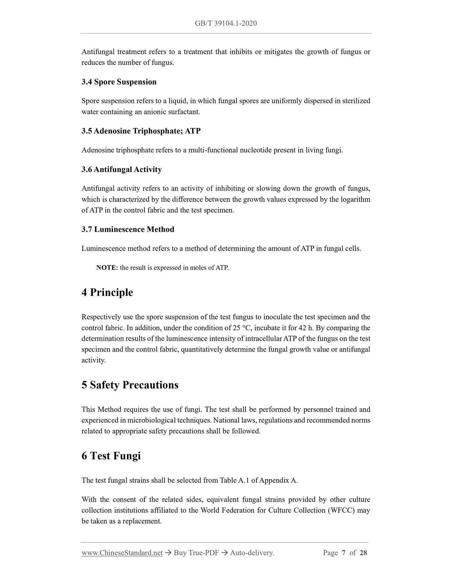
Antifungal treatment refers to a treatment that inhibits or mitigates the growth of fungus or
reduces the number of fungus.
3.4 Spore Suspension
Spore suspension refers to a liquid, in which fungal spores are uniformly dispersed in sterilized
water containing an anionic surfactant.
3.5 Adenosine Triphosphate; ATP
Adenosine triphosphate refers to a multi-functional nucleotide present in living fungi.
3.6 Antifungal Activity
Antifungal activity refers to an activity of inhibiting or slowing down the growth of fungus,
which is characterized by the difference between the growth values expressed by the logarithm
of ATP in the control fabric and the test specimen.
3.7 Luminescence Method
Luminescence method refers to a method of determining the amount of ATP in fungal cells.
NOTE: the result is expressed in moles of ATP.
4 Principle
Respectively use the spore suspension of the test fungus to inoculate the test specimen and the
control fabric. In addition, under the condition of 25 C, incubate it for 42 h. By comparing the
determination results of the luminescence intensity of intracellular ATP of the fungus on the test
specimen and the control fabric, quantitatively determine the fungal growth value or antifungal
activity.
5 Safety Precautions
This Method requires the use of fungi. The test shall be performed by personnel trained and
experienced in microbiological techniques. National laws, regulations and recommended norms
related to appropriate safety precautions shall be followed.
6 Test Fungi
The test fungal strains shall be selected from Table A.1 of Appendix A.
With the consent of the related sides, equivalent fungal strains provided by other culture
collection institutions affiliated to the World Federation for Culture Collection (WFCC) may
be taken as a replacement.
7.21 Microscope: with a magnification of 200.
7.22 Ultrasonic cleaner: the compact type for experiments, with a frequency of approximately
30 kHz ~ 50 kHz.
7.23 Luminometer: the test wavelength is 300 nm ~ 650 nm. Under the analytical conditions
specified in 8.4 and Chapter 11, it is capable of ATP measurement at the concentration of 1
10-8 mol/L ~ 1 10-5 mol/L.
7.24 pH meter: at 25 C, the accuracy is 0.1.
7.25 Refrigerator: capable of maintaining the temperature at 2 C ~ 10 C.
7.26 Freezers: one capable of adjusting the temperature to below 80 C, and the other capable
of adjusting the temperature to below 20 C.
Test tubes, glass vials, flasks, pipettes and tweezers shall be carefully washed with an alkaline
or neutral detergent, rinsed and dried; before use, they shall be processed through dry
sterilization or high-pressure steam sterilization.
8 Reagents and Culture Media
8.1 General
The reagents used for the test shall be analytically pure and / or suitable for microbiological
testing.
The existing commercial dehydration products should be used to prepare the culture media. In
addition, the instructions for use provided by the manufacturers of relevant products should be
strictly followed.
8.2 Pure Water
Analytical-grade pure water used for the preparation of microbial culture media and reagents.
It shall be obtained through distillation, ion exchange, ultrafiltration and / or reverse osmosis
(RO) device filtration; it shall be free from toxic or fungus-inhibitory substances.
8.3 Anionic Surfactant
Dioctyl sodium sulfosuccinate, used to prepare the spore suspension.
8.4 Luminescent Reagents, Reagents and Buffer Solutions
8.4.1 General
In accordance with the stipulations of 8.4.2 ~ 8.4.8, prepare reagents and buffer solutions. They
may be replaced by appropriately validated commercial reagents.
---Use the Pasteur pipette (7.13) or an equivalent device to pipette 0.5 mL of the sterilized
water containing anionic surfactant (8.4.8) (Step 1);
---By 5 times, disperse it into the spores in the center of the agar plate; slowly wash the
surface (Step 2).
NOTE: minor adjustments are acceptable, for example, increasing the amount of washing water.
Under this circumstance, keep records of all conditions.
10.3 Collection and Dispersion of Spore Suspension from a Culture Medium
See the steps below:
---Use the Pasteur pipette (7.13) or an equivalent device to pipette the spore suspension in
10.2;
---Transfer it into about 5 mL of the sterilized water containing anionic surfactant (8.4.8);
---Pipette about 100 times, or use a test tube agitator to agitate it, or perform light ultrasonic
cleaning for 5 min, so that the spores can be thoroughly dispersed;
---Visually observe that the suspension appears slightly cloudy (Step 3).
10.4 Filtering to Remove Hyphae and Spore Threads
Use a funnel or other devices with gauze (7.1) or glass wool to perform the filtration (Step 4).
NOTE: the size of the gauze (7.1) or glass wool can be 5 cm 5 cm, which is laid 4 layers 1
layer.
10.5 Use Centrifugal Separation and Re-suspension to Remove Supernatant
See the steps below:
---After filtration, under the condition of 25 C 2 C, at the centrifugal acceleration of
2,000 g, centrifuge for 5 min. When the centrifugal separator (7.18) does not have a
temperature control device, the centrifugation may be performed at room temperature;
---Remove the supernatant;
---Add 5 mL of the sterilized water containing anionic surfactant (8.4.8);
---Thoroughly pipette to disperse the spores, or use a test tube agitator to agitate it, or
perform light ultrasonic cleaning for 5 min, so that the spores can be thoroughly
dispersed (Step 5).
10.6 Confirmation of Concentration of Spore Suspension
Use a hemocytometer to check the following items:
a) The count and status of spores: confirm that the spore count is 1 106 CFU/mL ~ 3
106 CFU/mL, and over 90% of the spores are single spores free from hyphae;
b) When there are too many spores: use the sterilized water containing anionic surfactant
(8.4.8) to dilute the suspension, so that the spore count can reduce to 1 106 CFU/mL
~ 3 106 CFU/mL; check the spore count again;
c) When there are not enough spores: repeat the centrifugal separation to remove the
supernatant; use the sterilized water containing anionic surfactant (8.4.8) to adjust the
spore count to 1 106 CFU/mL ~ 3 106 CFU/mL; check the spore count again;
d) For the transfer method in 12.1.3, the spore count shall be adjusted to 1 108 CFU/mL
~ 3 108 CFU/mL. The spore suspension of this concentration shall be used for testing.
10.7 Adjustment of Spore Suspension for Testing
See the steps below:
---Use an anionic surfactant solution containing 5% SDB to adjust the concentration of the
spore suspension to 1 105 CFU/mL ~ 3 105 CFU/mL; reserve it for tests;
NOTE: for example, to prepare 100 mL of anionic surfactant solution containing 5% SDB: in 95
mL of the sterilized water containing anionic surfactant prepared in accordance with 8.4.8,
add 5 mL of the SDB culture medium prepared in accordance with 8.5.2.
---When diluting, thoroughly agitate the spore suspension;
---Under the condition of 3 C ~ 4 C, refrigerate the spore suspension and use it within 4
h.
11 Preparation of ATP Calibration Curve
The process of preparing the ATP calibration curve is as follows (the curve shall be drawn on
the day of the test):
a) Use pure water to dilute the ATP standard stock solution in 8.4.2, so as to prepare 3
dilutions with the exact concentrations of 1 108 mol/L, 1 107 mol/L and 1 106
mol/L;
b) In accordance with the following steps, prepare the first ATP dilution sample: transfer
0.1 mL from each dilution to a different plastic test tube, then, add 0.05 mL of pure
water and 0.35 mL of physiological saline solution to each plastic test tube and shake
it well;
c) In accordance with the following steps, prepare the second ATP dilution sample: take
0.1 mL from the first ATP dilution sample and add it to the test tube; add 0.4 mL of
physiological saline solution and shake it well;
Where,
Y---the ATP concentration, expressed in (mol/L);
X---the luminescence intensity (RLU is the relative light unit).
When the correlation coefficient between the mean luminescence intensity and
ATP concentration is lower than 0.99, the ATP calibration curve shall be re-drawn.
12 Test Methods
12.1 Specimen Preparation and Inoculation
12.1.1 General
The absorption method and transfer method can be adopted for the inoculation. The transfer
method is suitable for non-absorbent textiles.
12.1.2 Absorption method
12.1.2.1 Sample washing
If necessary, in accordance with method 10.1 of GB/T 20944.2-2007, wash the specimen. After
washing, use water to rinse the specimen, so as to remove the detergent. If other methods not
mentioned here are adopted, they shall be stated in the report.
12.1.2.2 Shape and mass of specimen
Cut a representative specimen from the sample and cut it into an appropriate size. Weigh-take
0.20 g 0.03 g as a test specimen. Respectively take six control fabrics and six test specimens.
NOTE: three of the control fabrics and three of the test specimens are used to determine the
luminescence intensity at the “0” contact time at the end of the inoculation. The remaining
sample is used to determine the luminescence intensity at the contact time after the
inoculation.
12.1.2.3 Placement of specimens
In accordance with the characteristic of the specimen, select one of the following methods and
respectively put each specimen into a different screw-top glass vial (7.9):
a) If the specimen is a textile that easily curls, or contains wadding or down, place a
glass rod on the specimen in the screw-top glass vial (7.9), or use thread to fix both
sides of the specimen;
b) In case of a yarn specimen, arrange the yarn into a bundle and place a glass rod on the
specimen in the screw-top glass vial (7.9);
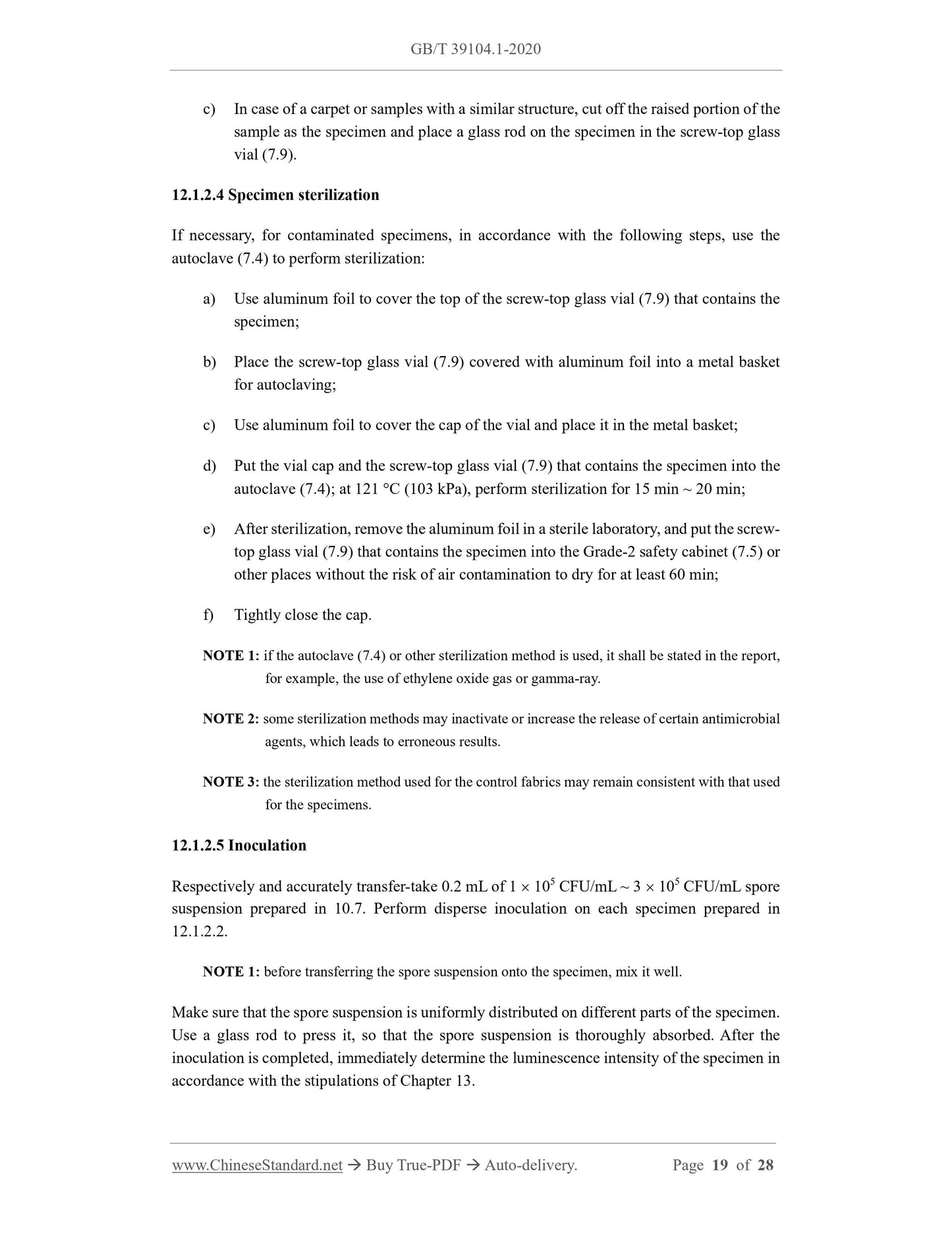
c) In case of a carpet or samples with a similar structure, cut off the raised portion of the
sample as the specimen and place a glass rod on the specimen in the screw-top glass
vial (7.9).
12.1.2.4 Specimen sterilization
If necessary, for contaminated specimens, in accordance with the following steps, use the
autoclave (7.4) to perform sterilization:
a) Use aluminum foil to cover the top of the screw-top glass vial (7.9) that contains the
specimen;
b) Place the screw-top glass vial (7.9) covered with aluminum foil into a metal basket
for autoclaving;
c) Use aluminum foil to cover the cap of the vial and place it in the metal basket;
d) Put the vial cap and the screw-top glass vial (7.9) that contains the specimen into the
autoclave (7.4); at 121 C (103 kPa), perform sterilization for 15 min ~ 20 min;
e) After sterilization, remove the aluminum foil in a sterile laboratory, and put the screw-
top glass vial (7.9) that contains the specimen into the Grade-2 safety cabinet (7.5) or
other places without the risk of air contamination to dry for at least 60 min;
f) Tightly close the cap.
NOTE 1: if the autoclave (7.4) or other sterilization method is used, it shall be stated in the report,
for example, the use of ethylene oxide gas or gamma-ray.
NOTE 2: some ster...
Share