1
/
of
5
www.ChineseStandard.us -- Field Test Asia Pte. Ltd.
GB/T 16886.5-2017 English PDF (GB/T16886.5-2017)
Regular price
$455.00
Regular price
Sale price
$455.00
Unit price
/
per
Shipping calculated at checkout.
Couldn't load pickup availability
GB/T 16886.5-2017: Biological evaluation of medical devices -- Part 5: Tests for in vitro cytotoxicity
Delivery: 9 seconds. Download (& Email) true-PDF + Invoice.
Get Quotation: Click GB/T 16886.5-2017 (Self-service in 1-minute)
Historical versions (Master-website): GB/T 16886.5-2017
Preview True-PDF (Reload/Scroll-down if blank)
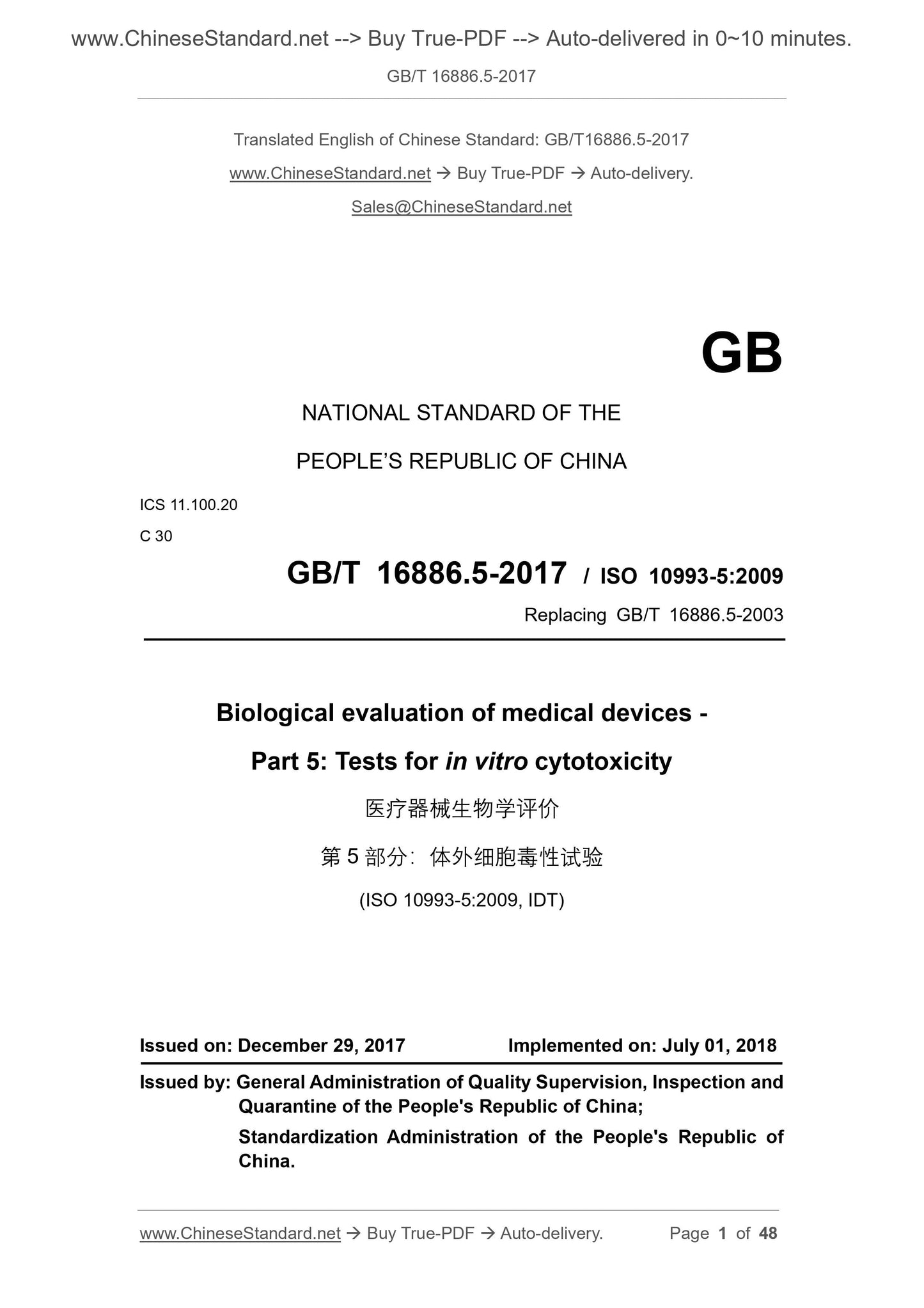
GB/T 16886.5-2017
Biological evaluation of medical devices--Part 5. Tests for in vitro cytotoxicity
ICS 11.100.20
C30
National Standards of People's Republic of China
Replace GB/T 16886.5-2003
Medical device biology evaluation
Part 5. In vitro cytotoxicity test
Part 5. Testsforinvitrocytotoxicity
(ISO 10993-5.2009, IDT)
Released on.2017-12-29
2018-07-01 implementation
General Administration of Quality Supervision, Inspection and Quarantine of the People's Republic of China
China National Standardization Administration issued
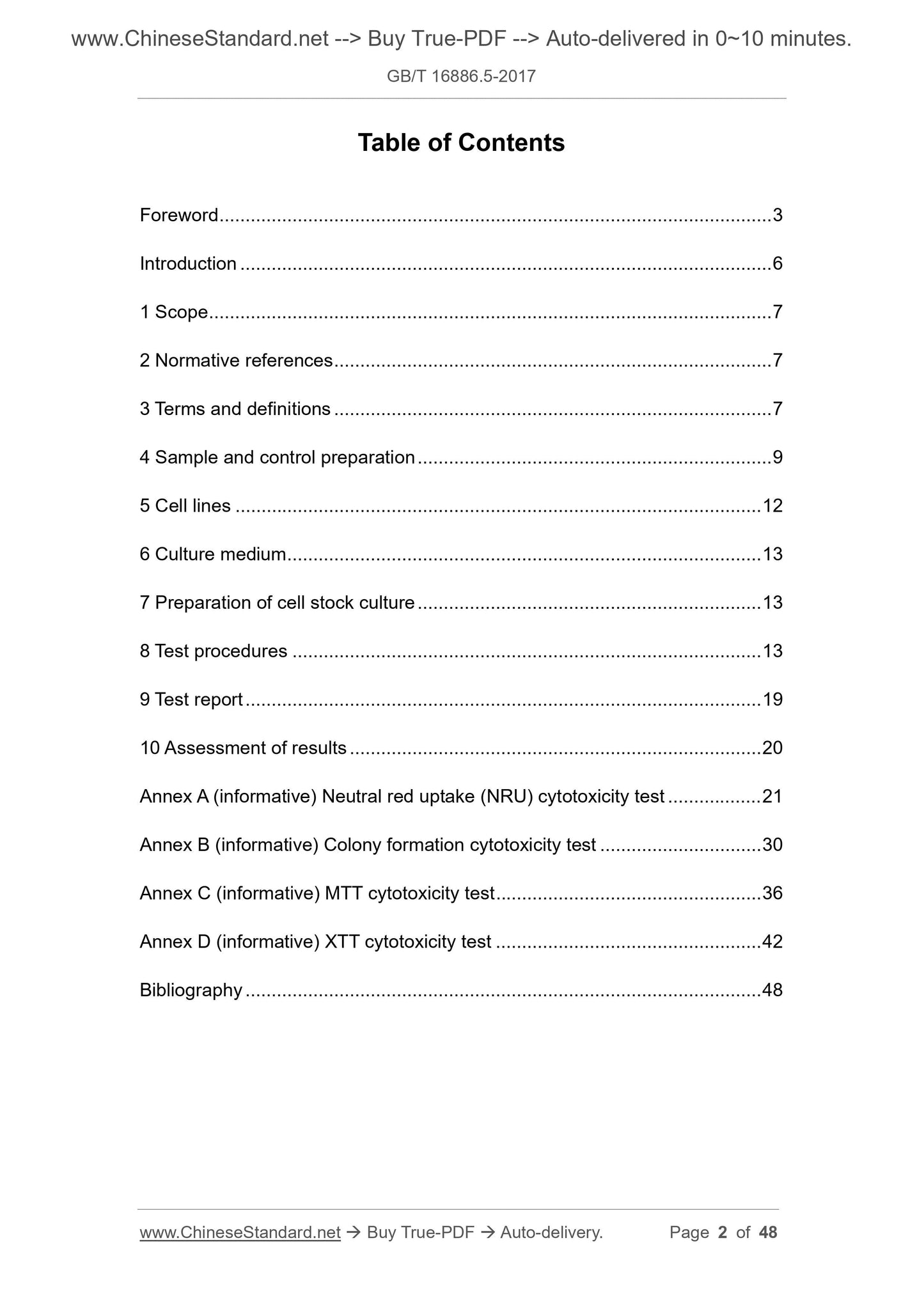
Content
Foreword III
Introduction V
1 range 1
2 Normative references 1
3 Terms and Definitions 1
4 sample and reference preparation 2
5 cell line 4
6 medium 4
7 Storage culture cell preparation 4
8 Test step 5
9 Test report 8
10 result evaluation 8
Appendix A (informative) Neutral red uptake (NRU) cytotoxicity test 9
Appendix B (informative appendix) Colony formation cytotoxicity test 15
Appendix C (informative) MTT cytotoxicity test 19
Appendix D (informative) XTT Cytotoxicity Test 23
Reference 27
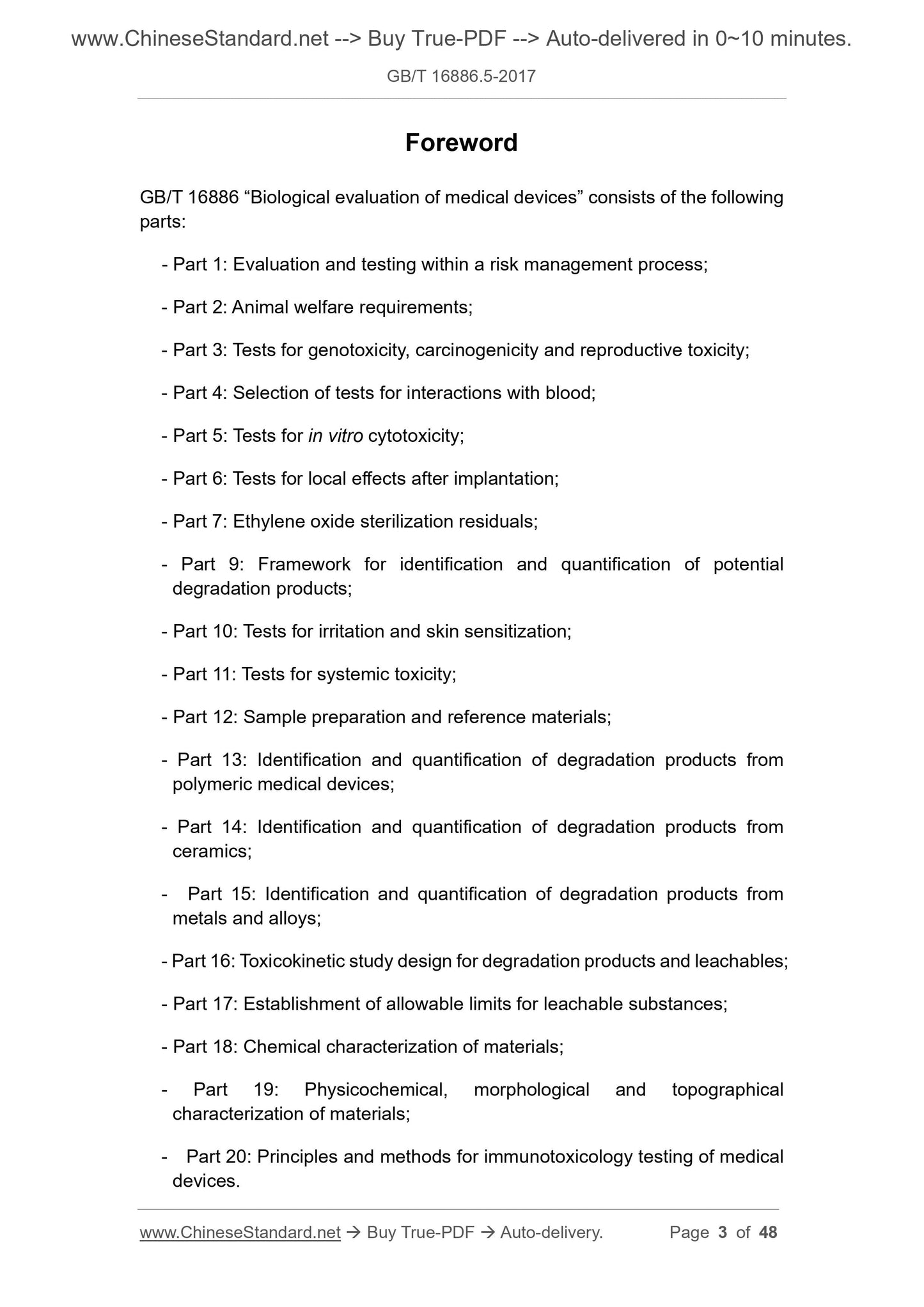
Foreword
GB/T 16886 "Biological Evaluation of Medical Devices" consists of the following components.
--- Part 1. Evaluation and testing in the risk management process;
--- Part 2. Animal welfare requirements;
---Part 3. Tests for genotoxicity, carcinogenicity and reproductive toxicity;
--- Part 4. Test options for interaction with blood;
---Part 5. In vitro cytotoxicity test;
--- Part 6. Post-implantation local reaction test;
---Part 7. Ethylene oxide sterilization residue;
---Part 9. Qualitative and quantitative frameworks for potential degradation products;
--- Part 10. Stimulation and delayed type hypersensitivity test;
--- Part 11. Systemic toxicity test;
--- Part 12. Sample preparation and reference materials;
--- Part 13. Qualitative and quantitative determination of polymer degradation products;
--- Part 14. Qualitative and quantitative determination of ceramic degradation products;
---Part 15. Qualitative and quantitative determination of metal and alloy degradation products;
---Part 16. Design of toxicokinetic studies of degradation products and leachables;
--- Part 17. The establishment of a limitable amount of leachables;
---Part 18. Chemical characterization of materials;
--- Part 19. Physicochemical, Morphological and Surface Characterization of Materials;
--- Part 20. Principles and methods for immunological testing of medical devices.
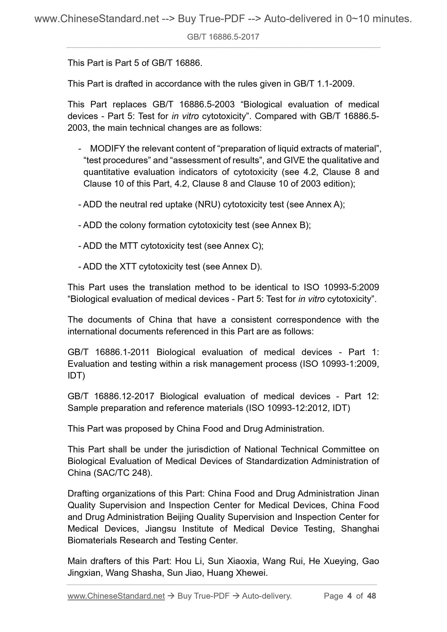
This part is the fifth part of GB/T 16886.
This part is drafted in accordance with the rules given in GB/T 1.1-2009.
This part replaces GB/T 16886.5-2003 "Biological evaluation of medical devices Part 5. In vitro cytotoxicity test", and
Compared with GB/T 16886.5-2003, the main technical changes are as follows.
--- Revised the "preparation of material extract", "test procedure", "result evaluation" and other related content, giving cytotoxicity qualitative and quantitative
Evaluation indicators (see 4.2, Chapters 8 and 10,.2003 editions 4.2, 8 and 10);
--- Increased sexual red intake (NRU) cytotoxicity test (see Appendix A);
--- Increased colony formation cytotoxicity test (see Appendix B);
--- Increased MTT cytotoxicity test (see Appendix C);
--- Increased XTT cytotoxicity test (see Appendix D).
This section uses the translation method equivalent to ISO 10993-5.2009 "Medical evaluation of medical devices Part 5. In vitro cytotoxicity
test".
The documents of our country that have a consistent correspondence with the international documents referenced in this part are as follows.
GB/T 16886.1-2011 Biological evaluation of medical devices - Part 1. Evaluation and testing in the process of risk management
(ISO 10993-1.2009, IDT)
GB/T 16886.12-2017 Biological evaluation of medical devices - Part 12. Sample preparation and reference materials (ISO 10993-12.
2012, IDT)
This part is proposed by the State Food and Drug Administration.
This part is under the jurisdiction of the National Technical Committee for Standardization of Medical Device Biology Evaluation (SAC/TC248).
This section drafted by. State Food and Drug Administration Jinan Medical Device Quality Supervision and Inspection Center, National Food and Drug Administration
Beijing Medical Device Quality Supervision and Inspection Center, Jiangsu Medical Device Inspection Institute, Shanghai Biomaterials Research and Testing Center.
The main drafters of this section. Hou Li, Sun Xiaoxia, Wang Rui, He Xueying, Gao Jingxian, Wang Shasha, Sun Wei, Huang Wei.
The previous versions of the standards replaced by this section are.
---GB/T 16886.5-1997;
---GB/T 16886.5-2003.
introduction
The in vitro cytotoxicity test is versatile and is widely applicable to the evaluation of various medical devices and materials. Therefore, the GB/T 16886 version
Part of the purpose is not to specify a single test method, but to specify a test plan that needs to be judged in a series of test steps to
Select the most appropriate test.
The tests are divided into three categories. extract test, direct contact test, and indirect contact test.
One or more of these tests were selected based on the nature of the sample being evaluated, the site of use, and the characteristics of use.
The choice of test determines the method of preparation of the test sample, the preparation of the cultured cells, and the manner in which the cells are contacted with the sample or its extract.
At the end of the contact test, the cytotoxicity and toxicity were evaluated. This part of GB/T 16886 released the evaluation party
The choice of this strategy allows for a range of trials to be available, reflecting many of the ideas that advocate the in vitro biological testing community.
The large number of methods and endpoint assays used in cytotoxicity assays can be divided into the following types of assessments.
--- According to the morphological method to assess cell damage;
--- Determination of cell damage;
--- Determination of cell growth;
--- Determination of cellular metabolic properties.
In each of these four types, there are several methods to choose from, and the researcher should understand the classification of the test and its corresponding special skills.
In order to be comparable to other results of similar instruments or materials at the level within and between laboratories. Appendix
An example of a quantitative test method is given. This part of GB/T 16886 also gives a guide to the interpretation of the test results.
Medical device biology evaluation
Part 5. In vitro cytotoxicity test
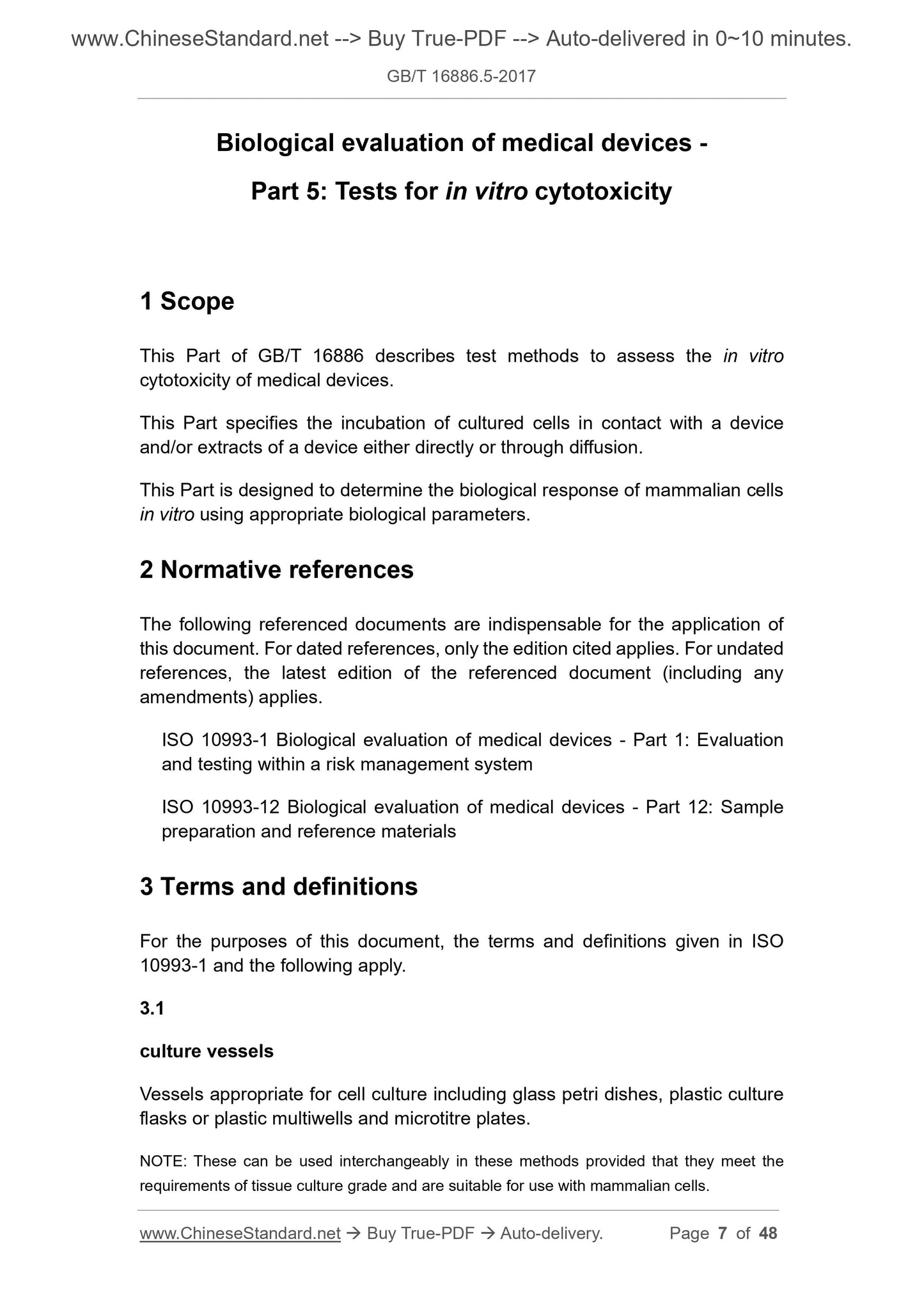
1 Scope
This part of GB/T 16886 describes test methods for assessing the in vitro cytotoxicity of medical devices.
This section specifies incubation methods for direct contact with the device and/or instrument extract or by contact with cultured cells by diffusion.
This section applies to the biological parameters of mammalian cells in vitro by suitable biological parameters.
2 Normative references
The following documents are indispensable for the application of this document. For dated references, only dated versions apply to this article.
Pieces. For undated references, the latest edition (including all amendments) applies to this document.
ISO 10993-1 Biological evaluation of medical devices - Part 1. Evaluation and testing in the process of risk management (Biologicalevalua-
tionofmedicaldevices-Part 1.Evaluationandtestingwithinariskmanagementprocess)
ISO 10993-12 Biological evaluation of medical devices - Part 12. Sample preparation and reference samples (Biologicalevaluationof
medicaldevices-Part 12. Samplepreparationandreferencematerials)
3 Terms and definitions
The following terms and definitions defined by ISO 10993-1 apply to this document.
3.1
Culture vessel culturevessels
Vessels suitable for cell culture, including glass culture dishes, plastic culture bottles or plastic porous culture plates and microtiter plates.
Note. In these test methods, these vessels are used interchangeably as long as they meet the requirements of tissue culture level and are suitable for mammalian cell culture.
3.2
Positive control material
Materials that reproduce cytotoxic reactions when tested in this section.
Note. The purpose of the positive control is to show the reaction of the applicable test system, such as polyurethane with organotin as a stabilizer 1) has been used as a solid material and leaching solution
For the control, a dilution of phenol was used as a positive control for the extract. In addition to one material, purified materials can be used to demonstrate the performance of the test system.
1) Polyurethane containing zinc diethyldithiocarbamate (ZDEC) and diethyldithiocarbamate (ZDBC) can be obtained from the Hatano Institute of Food Medicine
Product Safety Center (Ochiai729-5, Hadanoshi, Kanagawa257, Japan). The information given is for the convenience of users in this section, not
These products are approved on behalf of the standard publisher, and equivalent products can be used if they produce the same results.
3.3
Blank blank
The leaching medium containing no test sample is placed in the same vessel as the test sample during leaching and subjected to the same test sample
condition.
Note. The purpose of the blank is to evaluate the possible interference effects of the leaching vessel, the leaching medium and the leaching process.
3.4
Negative control material negativecontrolmaterial
Materials that do not produce cytotoxic reactions when tested in this section.
Note. The purpose of the negative control is to show the background reaction of the cells, such as high density polyethylene 2) as a negative control for synthetic polymers, and alumina ceramic rods.
Used as a negative control for dental materials.
2) High-density polyethylene is available from the US Pharmacopoeia Commission (Rockvile, MD, USA) and the Food and Drug Safety Center of the Hatano Institute (Ochiai729-
5, Hadanoshi, Kanagawa257, Japan) obtained. The information given is for the convenience of the users of this section and does not endorse the standard publisher.
For products, equal products can be used if they produce the same results.
3.5
Test sample testsample
A material, device, part of a device, component, extract, or a portion thereof used in biological testing, chemical testing, or evaluation.
3.6
Near convergence subconfluency
At the end of the logarithmic growth phase, approximately 80% of the cells meet.
4 sample and control preparation
4.1 General
The test should use.
a) test sample extract; and/or
b) Test the sample itself.
Sample preparation should be in accordance with ISO 10993-12.
Each test should include a negative control and a positive control.
4.2 Preparation of material extract
4.2.1 Principle of extraction
In order to determine potential toxicological hazards, the extraction conditions should be simulated or strictly in accordance with clinical conditions, unless required during clinical application.
However, it does not cause significant changes in the test material such as melting, dissolution or any change in chemical structure. Because of the properties of certain materials (such as
Degradable materials), chemical structure changes may occur during the extraction process.
Note. The concentration of any endogenous or exogenous substance in the extract and the amount of cells exposed to it depends on the interfacial area, extraction volume, pH, chemical dissolution.
Degree, diffusivity, osmotic pressure, agitation, temperature, time and other factors.
For devices (such as bone cement) where the patient mixes two or more components in use to become the final product, the device should not be advanced in advance.
Cleaning. Cleaning the test sample may reduce or remove residue from the device. If the test sample is used in a sterile environment,
The chemical components are leached using the sterilized test sample.
4.2.2 Leaching medium
The extraction medium is selected according to the chemical characteristics of the test sample and should be demonstrated and documented. Should be used in mammalian cell tests
List one or several media.
a) serum-containing medium;
b) a physiological saline solution;
c) Other suitable media.
The choice of medium should reflect the purpose of the leaching. Both polar and non-polar media should be considered. Serum-containing medium is the preferred dip
For medium extraction, serum-containing medium is preferred for leaching because it supports cell growth and leaching both polar and non-polar substances.
Ability. In addition to serum-containing media, serum-free media should be considered when it is clear that polar substances (such as ionic compounds) should be extracted.
Other suitable media include pure water and dimethyl sulfoxide (DMSO). In the selected test system, DMSO is higher than 0.5% (volume)
Score) is cytotoxic. Compared with the serum-containing medium extraction method, due to the dilution of the DMSO extract, the cells of the extractable cells are connected.
The touch concentration will be lower.
Note 1. Different types of serum (such as fetal bovine serum, bovine/fecal serum, newborn calf serum) may be used in the trial. Serum selection depends on cell type.
And set.
Note 2. It is important to consider that serum/protein is known to bind to the dissolution to some extent.
4.2.3 Extraction conditions
4.2.3.1 Leaching shall be carried out in a sterile, chemically inert closed container using aseptic technique and shall comply with ISO 10993-12.
4.2.3.2 Leaching shall be carried out in accordance with one of the following conditions and in accordance with the characteristics of the device and the specific use, except as given below.
a) (37 ± 1) ° C, (24 ± 2) h;
b) (50 ± 2) ° C, (72 ± 2) h;
c) (70 ± 2) ° C, (24 ± 2) h;
d) (121 ± 2) ° C, (1 ± 0.2) h.
The above extraction conditions are based on historical data and have been used to provide a measure of potential hazards in the assessment of the risk of an instrument or material. Other can be used
Conditions (such as prolonging or shortening the leaching time at 37 ° C), simulating leaching in clinical use or providing an appropriate measure of potential hazard
Set, but sh...
Share