GB/T 14698-2002 English PDF (GBT14698-2002)
Regular price
$120.00 USD
Regular price
Sale price
$120.00 USD
Unit price
/
per
Delivery: 3 seconds. Download true-PDF + Invoice.Newer version: (Replacing this standard) GB/T 14698-2017
Get QUOTATION in 1-minute: Click GB/T 14698-2002
Historical versions: GB/T 14698-2002
Preview True-PDF (Reload/Scroll if blank)
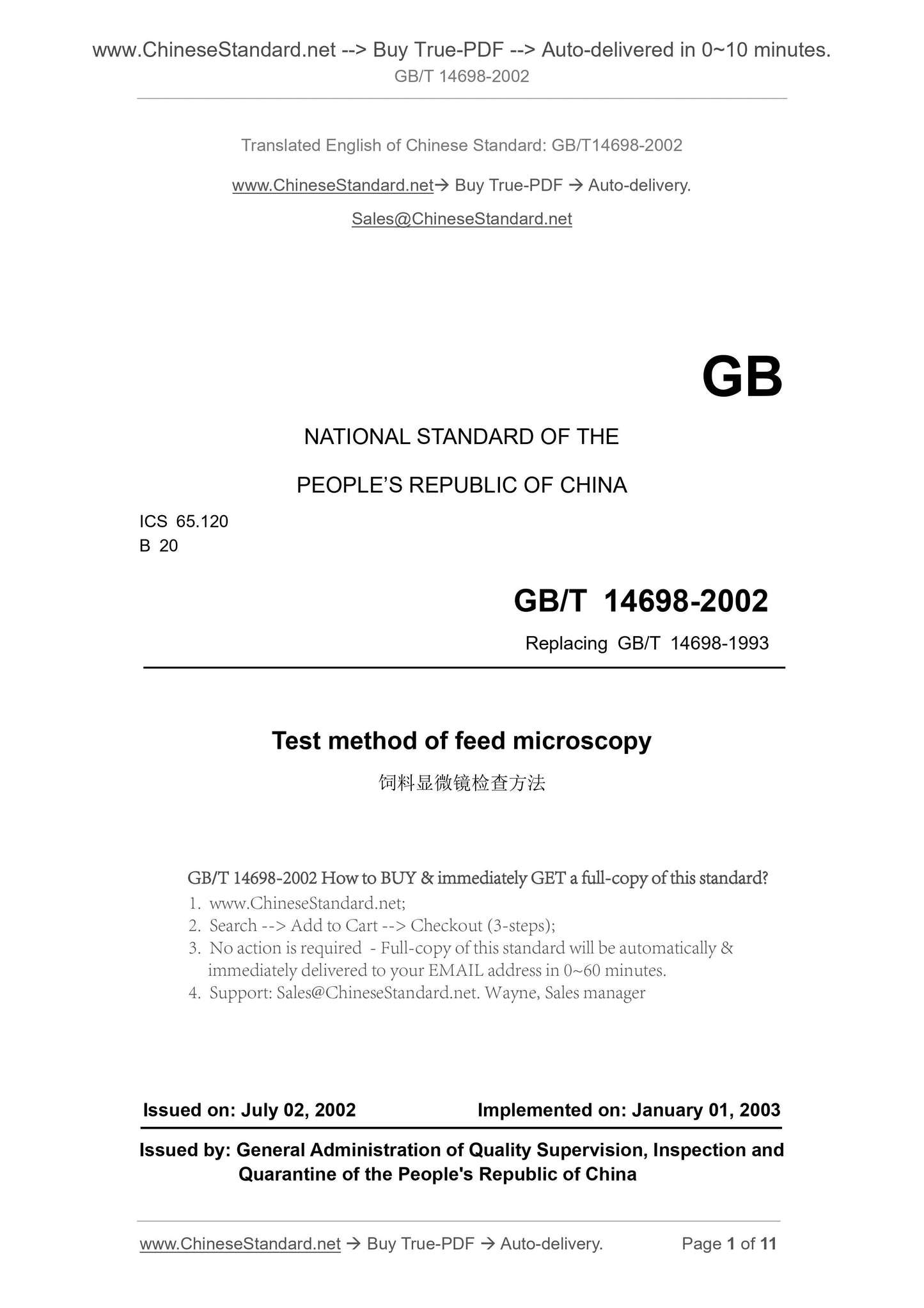
GB/T 14698-2002: Test method of feed microscopy
GB/T 14698-2002
GB
NATIONAL STANDARD OF THE
PEOPLE’S REPUBLIC OF CHINA
ICS 65.120
B 20
Replacing GB/T 14698-1993
Test method of feed microscopy
ISSUED ON. JULY 02, 2002
IMPLEMENTED ON. JANUARY 01, 2003
Issued by. General Administration of Quality Supervision, Inspection and
Quarantine of the People's Republic of China
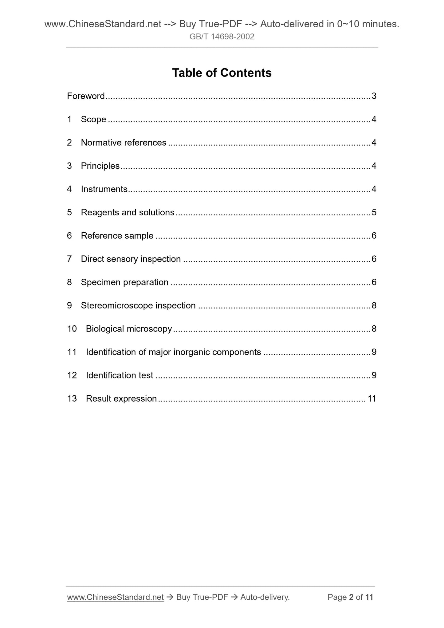
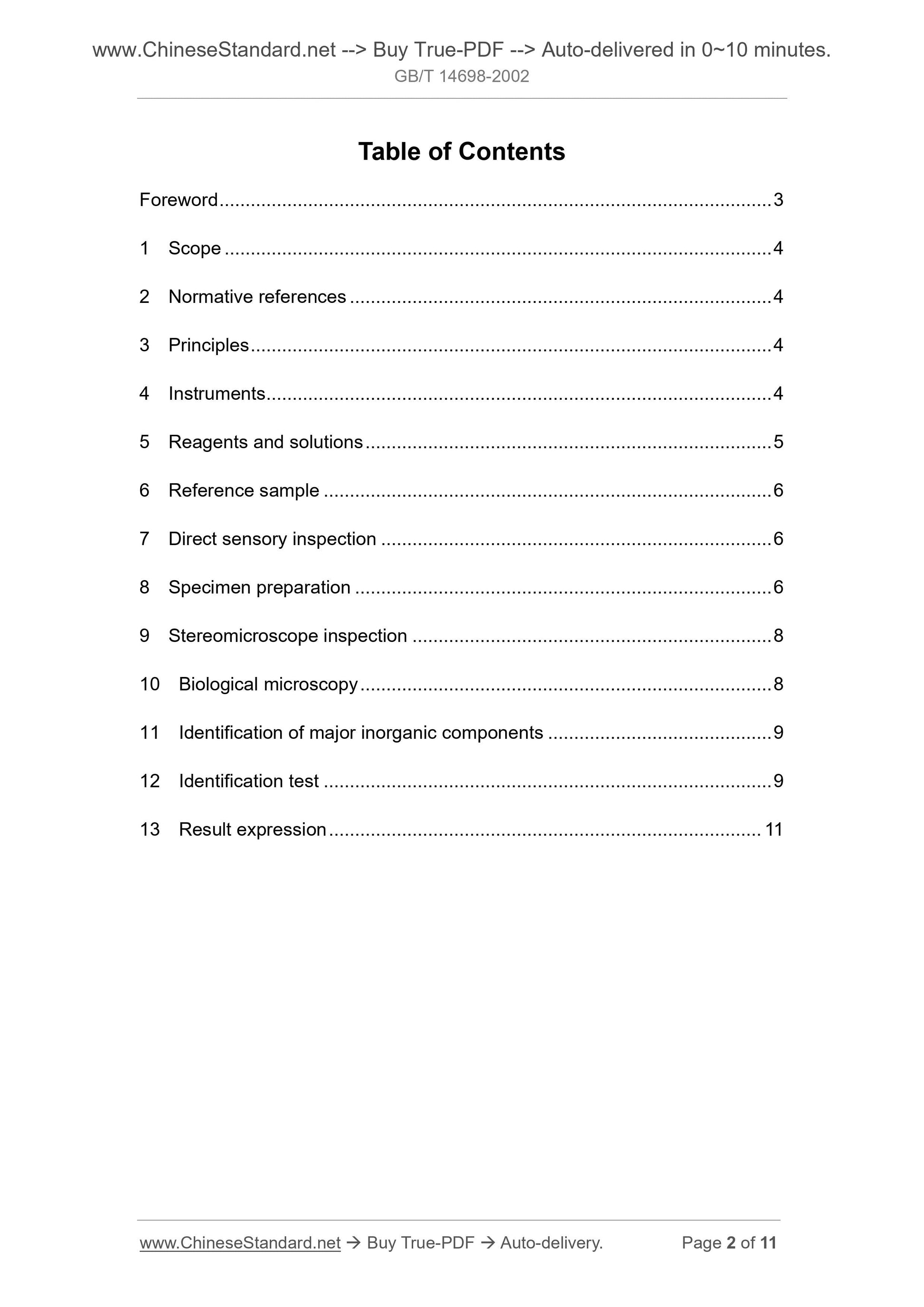
Table of Contents
Foreword ... 3
1 Scope ... 4
2 Normative references ... 4
3 Principles ... 4
4 Instruments ... 4
5 Reagents and solutions ... 5
6 Reference sample ... 6
7 Direct sensory inspection ... 6
8 Specimen preparation ... 6
9 Stereomicroscope inspection ... 8
10 Biological microscopy ... 8
11 Identification of major inorganic components ... 9
12 Identification test ... 9
13 Result expression ... 11
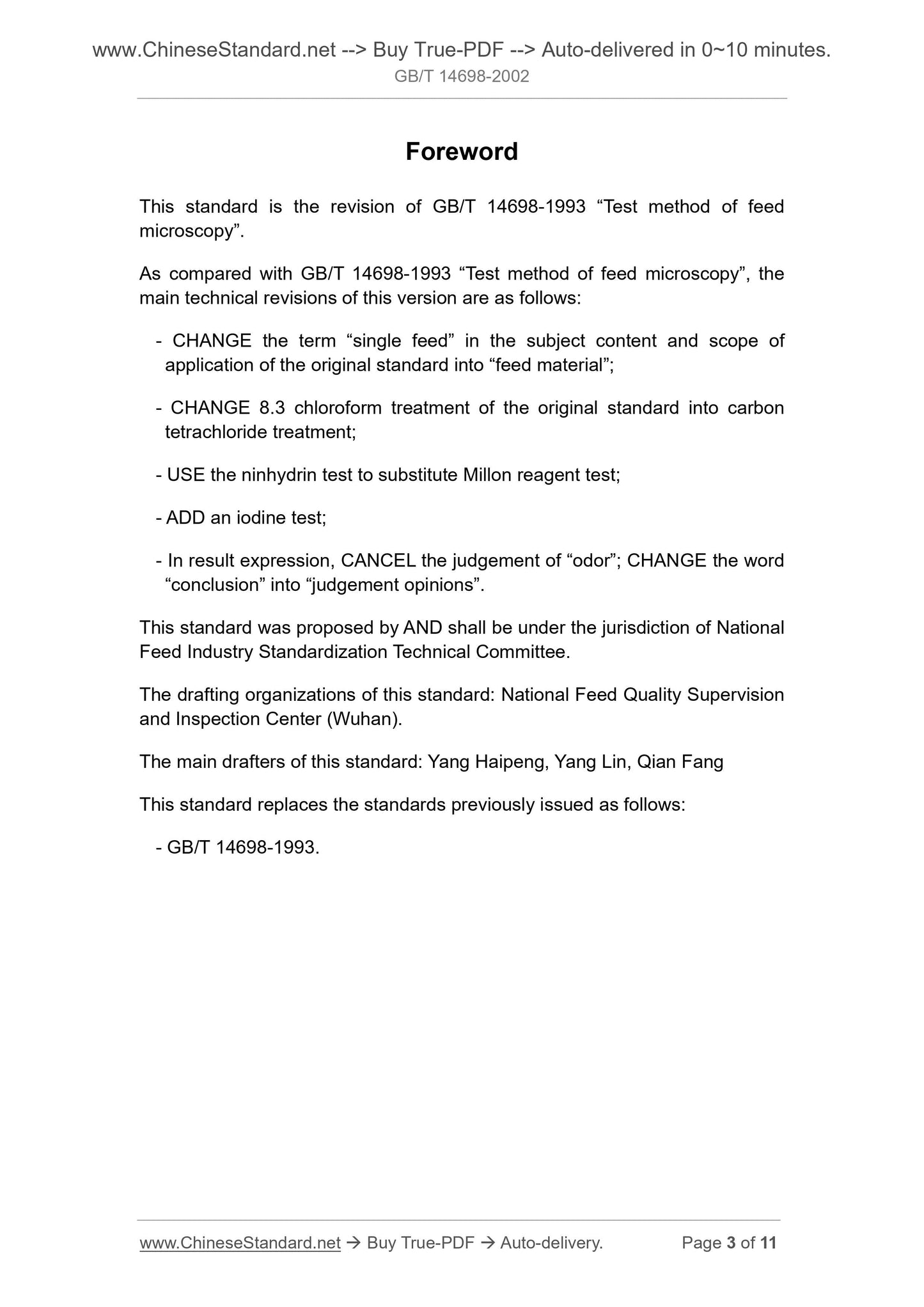
Foreword
This standard is the revision of GB/T 14698-1993 “Test method of feed
microscopy”.
As compared with GB/T 14698-1993 “Test method of feed microscopy”, the
main technical revisions of this version are as follows.
- CHANGE the term “single feed” in the subject content and scope of
application of the original standard into “feed material”;
- CHANGE 8.3 chloroform treatment of the original standard into carbon
tetrachloride treatment;
- USE the ninhydrin test to substitute Millon reagent test;
- ADD an iodine test;
- In result expression, CANCEL the judgement of “odor”; CHANGE the word
“conclusion” into “judgement opinions”.
This standard was proposed by AND shall be under the jurisdiction of National
Feed Industry Standardization Technical Committee.
The drafting organizations of this standard. National Feed Quality Supervision
and Inspection Center (Wuhan).
The main drafters of this standard. Yang Haipeng, Yang Lin, Qian Fang
This standard replaces the standards previously issued as follows.
- GB/T 14698-1993.
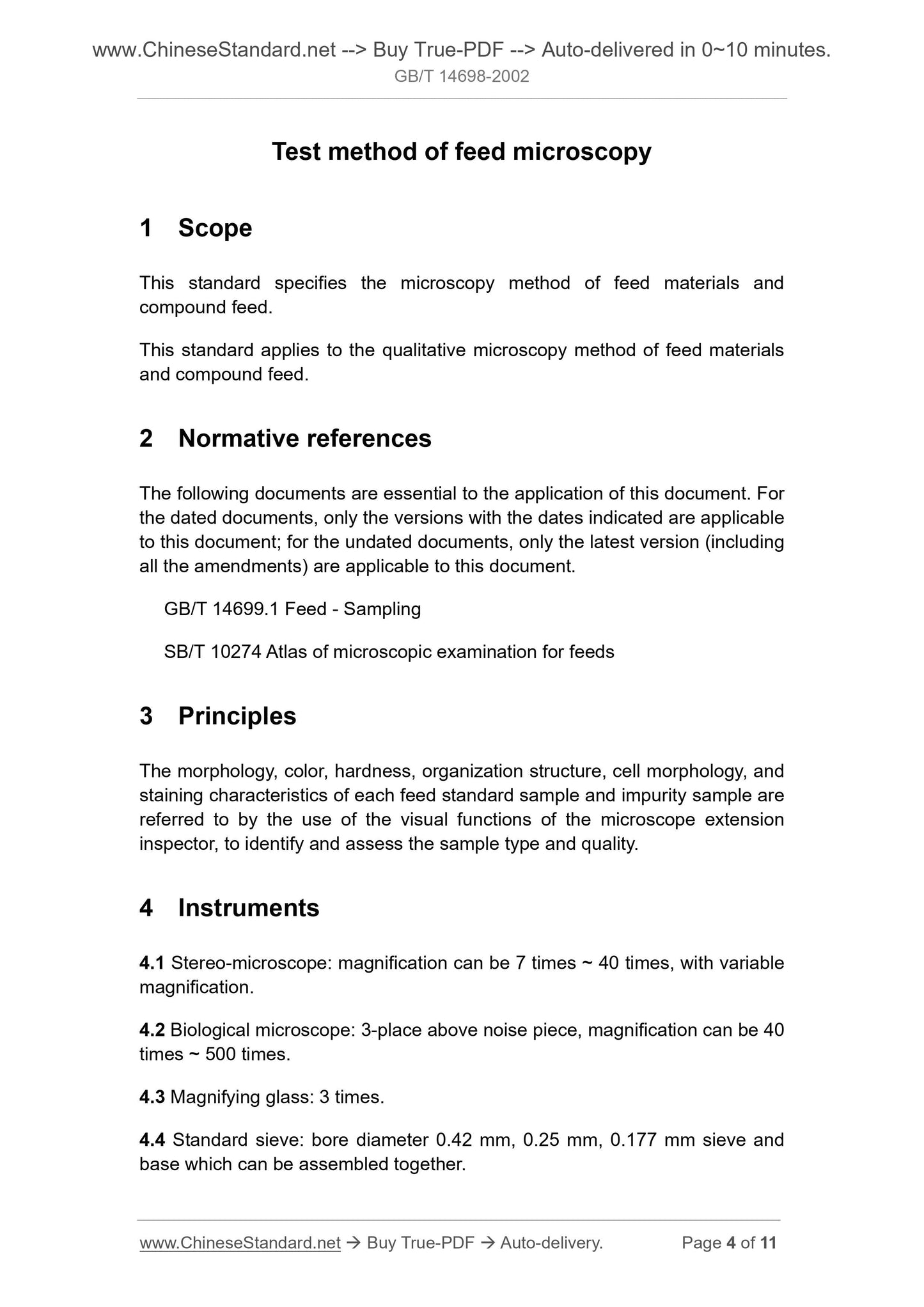
Test method of feed microscopy
1 Scope
This standard specifies the microscopy method of feed materials and
compound feed.
This standard applies to the qualitative microscopy method of feed materials
and compound feed.
2 Normative references
The following documents are essential to the application of this document. For
the dated documents, only the versions with the dates indicated are applicable
to this document; for the undated documents, only the latest version (including
all the amendments) are applicable to this document.
GB/T 14699.1 Feed - Sampling
SB/T 10274 Atlas of microscopic examination for feeds
3 Principles
The morphology, color, hardness, organization structure, cell morphology, and
staining characteristics of each feed standard sample and impurity sample are
referred to by the use of the visual functions of the microscope extension
inspector, to identify and assess the sample type and quality.
4 Instruments
4.1 Stereo-microscope. magnification can be 7 times ~ 40 times, with variable
magnification.
4.2 Biological microscope. 3-place above noise piece, magnification can be 40
times ~ 500 times.
4.3 Magnifying glass. 3 times.
4.4 Standard sieve. bore diameter 0.42 mm, 0.25 mm, 0.177 mm sieve and
base which can be assembled together.
TAKE sample in accordance with the feed sampling method in GB/T 14699.1,
MIX the specimen uniformly, USE the quartering method to reduce the sample
to the amount as required for inspection, generally 10 g ~ 15 g.
8.2 Screening
Based on the particle size of the specimen, SELECT the appropriate sieve
group, PLACE the sieve of the maximum bore diameter above the sieve of the
minimum bore diameter, PLACE the sieve base at the bottom. FULLY SHAKE
the specimen as taken by the quartering method on the sieve set, USE a spoon
to take some of specimens from above and below each sieve, respectively LAY
it horizontally in the culture dish [If necessary, the specimen can be subject to
carbon tetrachloride treatment first before being screened (as shown in 8.3)].
8.3 Carbon tetrachloride treatment
The specimen having higher fat content or attached with a large amount of fine
particles can be subject to carbon tetrachloride treatment first (fish meal, meat
and bone meal, most of poultry feed and unknown feed should be treated by
this method).
TAKE about 10 g of specimen, PLACE it into a 100 mL high beaker, ADD about
90 mL of carbon tetrachloride (5.1) (in the fume hood), STIR it for about 10 s,
LET it be standing for 2 min, after the upper and lower layers are separated
clearly, USE spoon to take out the floating substance, FILTER it, after it is
slightly evaporated dry, PLACE it in the oven at 70 °C for 20 min, TAKE it out to
cool it to room temperature, MAKE the specimen filtered. If necessary, it can
also filter, dry and screen the sediments.
8.4 Acetone treatment
For the specimen having a lump structure due to molasses OR having high
moisture content and being vague, it can be treated first using this method.
TAKE about 10 g of specimen, PLACE it into a 100 mL high beaker, ADD about
70 mL of acetone solution (5.2), STIR it for a few minutes to dissolve the
molasses, LET it be standing to allow it to settle. Carefully DECANT it, USE the
acetone solution to make repeated rinsing, settlement, and decantation for two
times. After it is slightly evaporated dry, PLACE it into a 60 °C oven for 20 min,
TAKE it out, COOL it at room temperature.
8.5 Particle or pellet specimen treatment
TAKE a few grains into a mortar, USE pestle to grind it to scatter it into different
compositions, BUT do not CRUSH the composition itself. After initial grinding,
MAKE it pass the sieve of bore diameter 0.42 mm. In accordance with the
further for each target. COMPARE it with the reference sample. TAKE off the
glass slide, LIFT up the cover slip, ADD one drop of iodine solution (5.5), STIR
it uniformly, ADD the glass slip again, PLACE it under microscope for
observation. At this time, the starch is dyed blue to black, yeast and other
protein cells are yellow to brown. If sample transparency is too low to be
observed easily, it may take a small amount of specimen, ADD about 5 mL of
suspending agent II (5.10), MAKE it boil for 1 min, COOL it down, TAKE 1 ~ 2
drops of bottom sediment on the glass slide, COVER the glass slip to perform
microscopic inspection.
11 Identification of major inorganic components
PLACE the dried sediment (8.3) on a sieve set composed of sieves having hole-
diameters 0.42 mm, 0.25 mm, 0.177 mm and base-plates to sieve it,
respectively PLACE the sieved four parts into the culture dishes, USE the
stereomicroscope to inspect it (refer to 9), the bone and scale of animal and
fish as well as the shell of molluscs are generally easily identifiable. The salt is
usually cubic; the calcite in limestone is rhombohedral.
12 Identification test
USE the tweezers to place the unknown particles on the spot plate, gently
CRUSH it, PERFORM the rest operations under stereomicroscope,
SEPARATE particles from each other to make them have a spacing of 2.5 cm,
DRIP 1 drop of relevant reagent around each particle, USE the fine glass rod
to push it into the liquid, OBSERVE the change at interface.
12.1 Silver nitrate test
PUSH the unknown particles into the silver nitrate solution (5.11) to make
observation.
12.1.1 If white crystals are formed and slowly become larger, it is indicated that
the unknown particles are chloride.
12.1.2 If yellow crystals and yellow flaky pieces are formed, it is indicated that
the unknown particles are ...